Published online: 22 September 2009
© Société Internationale de Chirurgie 2009
Abstract
Background Abdominal wall hernia repair after open abdomen management represents a surgical challenge, particularly due to muscle tension and lateral retraction. This study was designed to propose the use of Botulinum Toxin Type A (BTA) before abdominal wall hernia repair. Methods A prospective study of patients with abdominal wall hernia after open abdomen management was under- taken between September 2007 and January 2009. Bilateral BTA application was performed under electromyographic guidance at the abdominal wall. Transverse abdominal wall defect measurement was practiced at weekly intervals: clinically, in the first two patients, and with CT scan in the following 10 patients. Surgical closure was scheduled if no further hernia defect reduction was noted. Patients were followed at monthly hospital visits.
Results In the first two patients, a hernia defect reduction of 50 and 47.2%, respectively, was documented by the third week after BTA application, with no further reduction. In the 10 patients under CT scan hernia defect measurement, when comparing the initial mean transverse defect measure and at 4 weeks after BTA application (13.85 ± 1.49 cm vs.
T. R. Ibarra-Hurtado C. M. Nuño-Guzmán
J. E. Echeagaray-Herrera
Department of General Surgery, Antiguo Hospital Civil de Guadalajara, Av. Circunvalación Dr. Atl No. 154. Col. Independencia, Guadalajara, Jalisco 44340, México e-mail: tomasibarra2005@yahoo.com.mx
E. Robles-Vélez
Department of Plastic and Reconstructive Surgery, Antiguo Hospital Civil de Guadalajara, Guadalajara, Jalisco, México
J. de Jesús González-Jaime
Department of Rehabilitation Medicine, Antiguo Hospital Civil de Guadalajara, Guadalajara, Jalisco, México
8.6 ± 2.07 cm), an overall mean reduction of 5.25 ± 2.32 cm was observed (p \ 0.001; 95% confidence interval, 3.59–6.91). Hernia repair was performed, with no recurrences at a mean follow-up of 9.08 months. Conclusions BTA application before abdominal wall hernia repair seems to be useful. The lateral muscles paralysis achieved and transverse hernia defect reduction allows a minimal tension closure. To our knowledge, this is the first report of BTA application before abdominal wall hernia reconstruction.
Introduction
Open abdomen surgery after laparotomy is a therapeutic alternative in conditions, such as abdominal sepsis or trauma. Resulting abdominal wall defects represent a surgical challenge due to lateral muscle retraction. Several techniques for such hernia repair have been described [1, 2]. Muscle relaxation secondary to flaccid paralysis after BTA injection is well documented. The duration of such effect is no more than 6 months [3]. Lateral abdominal wall muscular paralysis and consequent relaxation leading to a transverse hernia defect reduction might facilitate surgical reconstruction.
Materials and methods
A prospective study of patients with abdominal wall hernia after open abdomen management was undertaken between September 2007 and January 2009. All patients had a total abdominal midline hernia defect. Lateral abdominal wall musculature electromyography (EMG) was performed (XLTEK Electromyograph, XCALIBUR LT model. Bristol

Fig. 1 EMG-guided sites of BTA application on right flank (black dots) of one of the patients. BTA application was performed on the left flank under EMG-guidance as well
Circle, Oakville, Ontario, Canada.). A myoject needle (TECANeedles, MyoJect Disposable Hypodermic Needle Electrode, VIASYS Healthcare, Madison, WI, USA) was used to identify the maximal EMG recording points. Five points were identified at each side: two over the midaxillary line, between the costal border and the superior iliac crest, and three over the external oblique muscle (Fig. 1). Fifty units of BTA (Dysport, IPSEN, France) were applied through the myoject needle at each point (250 units at each side, for a total of 500 units per patient). In the first two patients, transverse abdominal wall defect clinical measurement was performed at weekly intervals. In the following ten cases, to document a more objective measurement of the abdominal wall defect, CT scan was per- formed before BTA application and 4 weeks after. Surgical closure was scheduled when no further defect reduction was documented. Postoperative follow-up was performed at monthly hospital visits.
Operative technique
Through an elliptical incision, scar tissue and previous incision were removed. Skin flaps were dissected laterally into both flanks. Hernia sac was dissected and excised completely. Entrance to peritoneal cavity and lysis of
adhesion between subjacent viscera and anterior abdominal wall was achieved. Midline rectus muscles fascial borders were identified bilaterally. Abdominal wall reconstruction was practiced with simple closure or components separa- tion [4]. Procedures for bowel continuity reestablishment were performed when indicated.
Descriptive statistics, including means and standard deviations, were used to describe the study population continuous variables. Differences between the initial and week-4 transverse hernia defect measures were assessed with a paired Student’s t test; p value \ 0.05 was defined as significant. Statistical analysis was performed using SPSS for Windows, version 12.0 (SSPS Inc., Chicago, IL).
Results
Twelve patients were included during study period. In the first two cases, an important reduction in the transverse defect measure was found after BTA application, with no further reduction at week 4 (Table 1). In the following ten cases under CT scan measurement, when comparing the initial mean transverse hernia defect measure and at 4 weeks after BTA application (13.85 ± 1.49 cm vs. 8.6 ± 2.07 cm), an overall mean reduction of 5.25 ± 2.32 cm was observed (p \ 0.001; 95% confidence interval (CI), 3.59–6.91). Simple closure technique was performed in six cases, and components separation tech- nique in the remaining six cases, with an overall compli- cation rate of 16.67%; a case of seroma formation managed under US-guided drainage, and a colocutaneous fistula, which closed with conservative treatment. No mortality was documented. After a mean follow-up of 9 months, no recurrences have been observed. Table 2 summarizes clinical and demographic characteristics of the ten cases with CT scan measurement. Figures 2 and 3 show the CT scans of case 3, before BTA application and 4 weeks after.
Discussion
Abdominal wall hernia reconstruction after open abdomen management represents a surgical challenge. The
Table 1 Demographic and clinical characteristics of patients with abdominal wall hernia after open abdomen management Transverse hernia defect weekly measures (cm)
Clinical transverse hernia defect measurements, initial and 4 weeks after BTA application Comp. separation Components separation
Table 2 Demographic and clinical characteristics of patients with abdominal wall hernia after open abdomen management Transverse hernia defect measures (cm)
CT scan transverse hernia defect measurements, initial and 4 weeks after BTA application * t = 7.14; 95% CI 3.59–6.91; p value \ 0.001, derived from paired Student’s t test
EC fistula enterocutaneous fistula, AP acute pancreatitis, AD acute diverticulitis, AA acute appendicitis, Comp. separation components sepa- ration, SD standard deviation, CI confidence interval
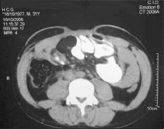
 Fig.2 Case4 CTscanprevioustoBTAapplication,showinga16.5- cm transverse hernia defect at mesogastric level
postoperative hernia rate after primary surgery is 2–20% [1], whereas the ventral hernia recurrence rate is reported to be 30–70% [1, 2, 5]. Surgical techniques consisting of myofascial release, local tissue flaps, free flaps, and the use of synthetic materials have been reported [6–12]. The ideal objective is to perform a tension-free closure, with abdominal wall dynamic stability and optimizing aesthetic appearance [13]. Planned ventral hernia is an alternative for seriously ill patients due to abdominal sepsis or trauma [12]. After a laparotomy, there is a disturbance of the abdominal wall dynamic forces. The midline is the most frequent site of post-incisional hernia formation.

Fig. 3 Case 4 CT scan 4 weeks after BTA application, showing a 6- cm transverse hernia defect
Although multifactorial, the most important cause is the lateral retraction and migration of midline structures. Lateral myofascial contraction develops a continued unopposed force, enhancing the hernia defect [14–16]. Several tech- niques consisting of myofascial release have been descri- bed [2–9, 12–15]. The subsequent myofascial relaxation provided by the lateral abdominal muscles, particularly the external oblique, allows midline rectus muscle approxi- mation and hernia defect closure with less tension. Cakmak et al. [17] reported the effect of BTA-induced abdominal muscle paralysis on intra-abdominal pressure in rats. In our series, BTA application produced a lateral abdominal wall
muscle relaxation, particularly over the external oblique muscles. Maximal relaxation developed at the third week of toxin application. BTA-induced flaccid paralysis con- tributed to a diminished transverse hernia defect and con- sequently to a less-tension abdominal wall reconstruction, encouraging the authors to report these results. During follow-up, no bulging over the oblique muscles has been observed. The ideal BTA dosage is to be determined. To our knowledge, this is the first report of BTA application before abdominal wall hernia reconstruction.
Conclusions
BTA application before abdominal wall hernia recon- struction seems to be useful. The lateral muscles paralysis achieved and a transverse hernia defect reduction allows a less tension closure.
Disclosure There was no external financial support for the achievement of this study. There is no conflict of interest to report by the authors.
References
- Cheng H, Rupprecht F, Jackson D et al (2007) Decision analysis model of incisional hernia after open abdominal surgery. Hernia 11:129–137
- Jacobs DG, Pratt BL, Capizzi PJ (2002) Reconstruction of the massive posttraumatic abdominal wall hernia. Prob Gen Surg 19:73–83
- Rohrer TE, Beer K (2005) Background to Botulinum toxin. In: Carruthers A, Carruthers J (eds) Botulinum toxin. Elsevier Saunders, Philadelphia, pp 9–17
- Ramirez OM, Ruas E, Dellon AL (1990) ‘‘Components separa- tion’’ method for closure of abdominal-wall defects: an anatomic and clinical study. Plast Reconstr Surg 86:519–526
- Helton WS, Fisichella PM, Berger R et al (2005) Short-term outcomes with small intestine submucosa for ventral abdominal hernia. Arch Surg 140:549–560
- Jacobsen WM, Petty PM, Bite U et al (1997) Massive abdominal wall hernia reconstruction with expanded external-internal obli- que and transversalis musculofascia. Plast Reconstr Surg 100:326–335
- Lindsey JT (2003) Abdominal wall partitioning (the accordion effect) for reconstruction of major defects: a retrospective review of 10 patients. Plast Reconstr Surg 112:477–485
- Jernigan TW, Fabian TC, Croce MA et al (2003) Staged man- agement of giant abdominal wall defects: acute and long-term results. Ann Surg 238:349–355
- Sleeman D, Sosa JL, Gonzalez A et al (1995) Reclosure of the open abdomen. J Am Coll Surg 180:200–204
- Shestak KC, Edington HJ, Johnson RR (2000) The separation of anatomic components technique for the reconstruction of massive midline abdominal wall defects: anatomy, surgical technique, applications, and limitations revisited. Plast Reconstr Surg 105:731–738
- Rohrich RJ, Lowe JB, Hackney FL et al (2000) An algorithm for abdominal wall reconstruction. Plast Reconstr Surg 105:202–216
- Fabian TC, Croce MA, Pritchard FE et al (1994) Planned ventral hernia. Staged management for acute abdominal wall defects. Ann Surg 219:643–650
- Mathes SJ, Steinwald PM, Foster RD et al (2000) Complex abdominal wall reconstruction: a comparison of flap and mesh closure. Ann Surg 232:586–596
- Levine JP, Karp NS (2001) Restoration of abdominal wall integrity as a salvage procedure in difficult recurrent abdominal wall hernias using a method of wide myofascial release. Plast Reconstr Surg 107:707–716
- Lowe JB, Garza JR, Bowman JL et al (2000) Endoscopically assisted ‘‘components separation’’ for closure of abdominal wall defects. Plast Reconstr Surg 105:720–729
- DuBay DA, Choi W, Urbanchek MG et al (2007) Incisional herniation induces decreased abdominal wall compliance via oblique muscle atrophy and fibrosis. Ann Surg 245:140–146
- Cakmak M, Caglayan F, Somuncu S et al (2006) Effect of paralysis of the abdominal wall muscles by botulinum A toxin to intraabdominal pressure: an experimental study. J Pediatr Surg 41:821–825
Maillot De Foot Femme
I really enjoy the blog.Much thanks again. Great.
Dr Tomas
Thanks to you for reading me It would be very nice and share my articles 😉
bayern munich shirt
It`s really useful! Thank you!
Dr Tomas
Thank you for your comments!